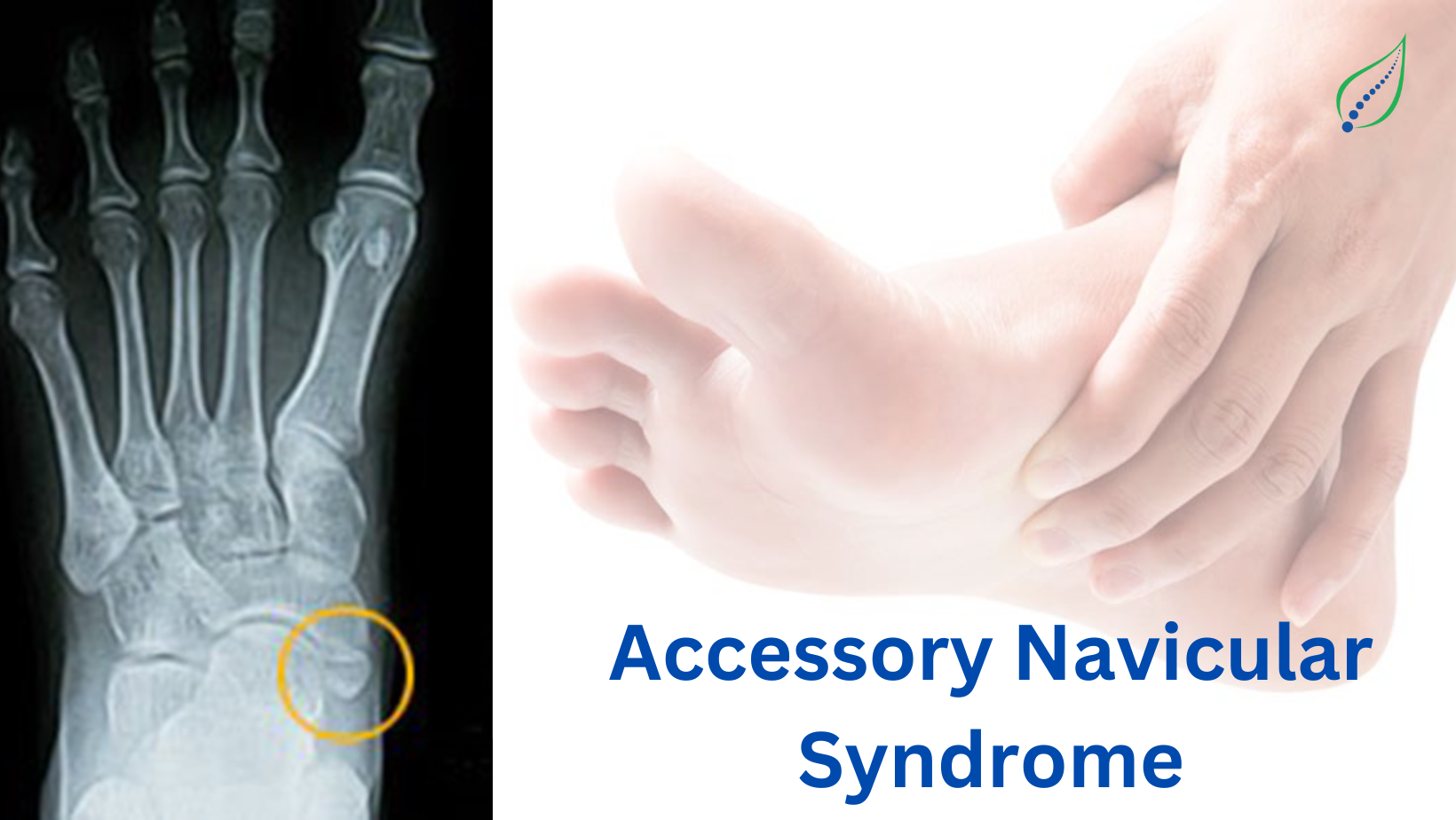
Accessory Navicular Bone
Accessory navicular syndrome is an extra bone or piece of cartilage located adjacent to the navicular on medial side of the foot. It presents as a sesamoid in the posterior tibial tendon.
Tendon of tibialis posterior inserts into the navicular bone. This muscle helps in ankle plantar flexion & plays a major role in supporting the medial arch of foot.
An accessory navicular bone lies posterior to posteromedial tuberosity of tarsal navicular. Presence of which compromises the suspension of posterior tibial tendon. It also causes posterior tibial tendinopathy as the insertion of the tibialis posterior tendon onto the accessory navicular is more proximal. Leverage of the malleolus on the tibialis posterior tendon is reduced increasing stress on the tendon. The calcaneal pitch angle is also reduced in patients with a symptomatic accessory navicular than in normal subjects.
It is classified into 3 types:
- Sesamoid bone in posterior tibial tendon
- Accessory bony growth 1.2cm in diameter, where synchondrosis develops between it and the navicular bone
- Fused accessory navicular resulting in large shaped navicular
A type I accessory navicular is rarely symptomatic. Where symptoms do appear, type I usually responds well to conservative management.
Patients with Type II have an increased risk of injury and onset is usually insidious or a result of trauma.
With Type III, excising the accessory navicular is indicated over excising the navicular prominence.
SYMPTOMS:
- Most of the time it is asymptomatic.
- Condition causes medial side foot pain.
- The pain is aggravated by walking, running and weight-bearing activities.
INVESTIGATIONS:
- XRAY
- MRI- Bone marrow oedema can be seen
- Gait examination
TREATMENT:
- Medications: Non steroidal anti-inflammatory drugs to reduce the pain.
- Rest & Icing: Immobilize the foot for 2-3 weeks & apply icing for 3-4 times/day on the painful site.
- Physical therapy: Start with gentle foot stretches & exercise to maintain ankle mobility. Exercise to strengthen the peroneal, tibialis posterior & intrinsic foot muscles.
- Well-padded shoe orthotics should be worn for arch support.
- Gait re-training, stability & balance exercises.
_1677139742.png)