Cervical Spinal Canal Stenosis
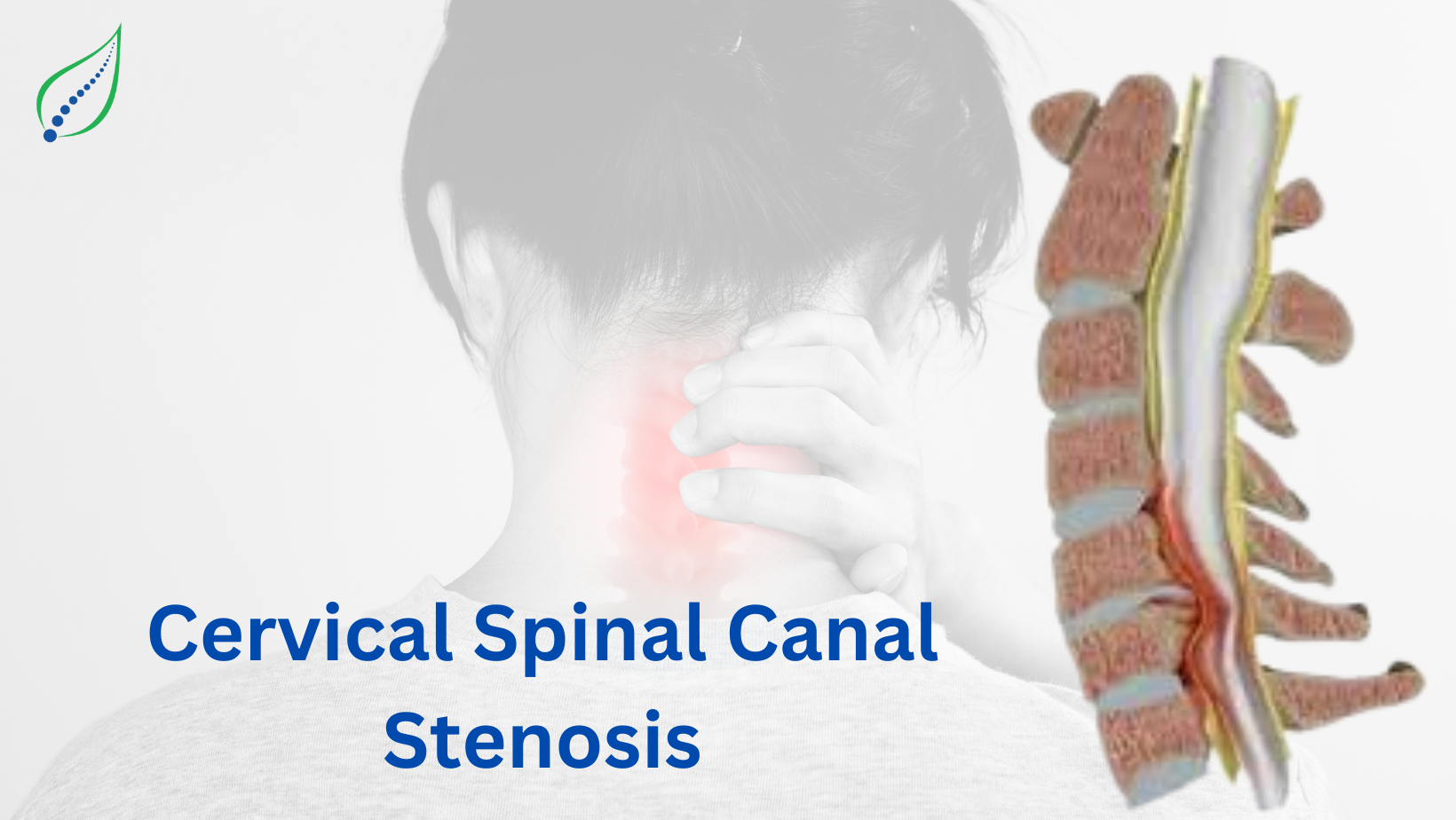
Cervical Spinal Canal Stenosis is signified by the narrowing of spinal canal in the neck region, which exerts pressure on the spinal cord and the nerve roots. If proper diagnosis and treatment is neglected, this compression could result in pain, numbness, and possible serious neurological issues. Although it is frequently observed in older individuals but depending on underlying conditions it can affect individuals of all ages.
The spinal canal is a hollow region inside the vertebrae containing the spinal cord and nerve roots. When this area narrows it decreases the amount of space accessible to the spinal cord, potentially resulting in compression and a number of symptoms. The severity of symptoms depends heavily upon the level of stenosis and whether the spinal cord or nerve roots are involved.
Symptoms
Symptoms of Cervical Spine Canal Stenosis is different based on the extent of narrowing as well as the number of nerves involved. Common symptoms include:
- Neck pain – This is the commonest symptom present in the patients with this condition. Mostly the pain is chronic may radiate to the shoulders or arms.
- Numbness and tingling – Most commonly felt in palms, arms, fingers or sometimes around the neck. Sometimes if patient has severe compression this also extends to legs
- Weakness – In case of severe compression of the spinal cord, it affects the corticospinal tract which is responsible for sending motor signals from brain to the muscles. Its disruption causes impaired voluntary muscle control leading to muscle weakness, spasticity and difficulty in performing motor tasks.
- Loss of coordination - Loss of coordination in cervical canal stenosis occurs due to spinal cord compression, disrupting motor control and proprioception pathways, leading to weakness, spasticity, gait disturbances, and impaired fine motor skills. Fine motor abilities may be impaired, creating challenges when performing daily tasks such as buttoning a shirt or grasping utensils.
- Bladder or bowel dysfunction - Severe cases can affect autonomic control, resulting in incontinence or difficulty with bowel movements. This factor plays a very important role in understanding whether patient can be treated non surgically or not.
- Headaches - Cervicogenic pain is a prevalent cause of headaches in cervical canal stenosis, in which spinal nerve compression and inflammation leads to the radiating pain to the head. This can also be caused by muscle tension and improper posture as patients tend to adjust their necks position to reduce pain, putting more strain on the upper neck muscles. In addition, due to reduced blood flow and nerve irritation may trigger headaches, particularly at the occipital area.
- Electric shock sensations – It is known as Lhermitte's sign. Lhermitte's sign is a typical symptom of cervical canal stenosis. When the neck is flexed forward, it causes a sudden, electric shock-like sensation that propagates down the spine and into the limbs. The event happens as a result of irritation or compression of the spinal cord dorsal columns, which are responsible for transferring sensory information. It is frequently related with cervical spinal cord diseases such as stenosis, multiple sclerosis, and B12 deficiency.
Symptoms generally improve over time, although an injury or rapid increase in compression might cause an acute aggravation.
Causes
Congenital spinal stenosis is a condition in which when a child is born with anatomical defects that narrow the spinal canal, increasing the possibility of spinal cord or nerve compression. Congenital stenosis, as compared to degenerative stenosis, develops at birth as a result of multiple genetic or developmental abnormalities. Here are the most common congenital causes:
Congenital Causes:
- Achondroplasia:
It’s a genetic bone growth condition that causes limbs to grow excessively short and causes dwarfism. It results from mutations in the FGFR3 gene, which impacts on the development of bones and cartilage development. Nerve compression is more likely in people with achondroplasia because their spinal canals are abnormally narrow, especially in the cervical and lumbar areas. - Spinal Dysraphism:
It is a group of developmental abnormalities characterized by improper function of spine, spinal cord, nerve roots or during fetal growth is known as spinal dysraphism.
One of the commonest examples is spina bifida, in which a part of the spinal cord and its covering extends outside the spinal canal.
- Congenital Kyphosis: A spinal deformity caused by abnormal vertebral growth during pregnancy, congenital kyphosis leads the upper back to arch excessively outward. It can often come on by poor segmentation or abnormalities in the spine such as hemivertebrae. Because of the smaller room inside the spinal canal caused by this excessive curvature, there is a greater chance of spinal cord compression. Back pain, limited mobility, and neurological impairments are possible symptoms for those who are affected.
- Congenital Short Pedicles: These are short growths of bone that connect each vertebra's anterior (vertebral body) and posterior (lamina). These pedicles that extend are shorter than typical in congenital circumstances which decreases the spinal canal's entire size.
- Osteopetrosis: It is also referred to as "marble bone disease," osteopetrosis is a rare genetic condition that causes extremely hard and dense bones. This condition is brought on by poor bone resorption, resulting in the spinal canal and foramina to shrink.
- Morquio Syndrome: This rare genetic condition damages the development of bone, cartilage, and the spine by interfering with the body's capacity to break down particular molecules. Cervical spinal stenosis, abnormal spine curvature, and a tiny trunk are common in people with this disorder.
- Hereditary Multiple Exostoses: (Diaphyseal Aclasis): The hereditary disorder is marked by the growth of multiple exostoses, or bone protrusions, along the vertebrae as well as additional bones. The spinal cord or nerve roots may be compressed as a result of these growths' ability to compress the spinal canal. Depending on the size and location of the growths, symptoms may include pain, restricted movement, and neurological problems.
Acquired Causes:
Cervical spinal stenosis can develop gradually as an outcome of a number of acquired conditions that cause the spinal canal to narrow. Unlike congenital stenosis, which is present from birth, acquired stenosis can be caused by degenerative changes, trauma, or spine-related disorders. The following are the primary acquired causes of cervical spinal stenosis.
Degenerative Disc Disease (DDD):
One of the major causes is Degenerative Disc Disease (DDD). As people age, the intervertebral discs that provides cushioning to the vertebrae lose water and shrink, reducing their height. The deterioration brings the vertebrae closer to one another, leading to spinal canal narrowing. In addition, disc degeneration may result in bulging or herniation, exerting pressure on the spinal cord and nerve roots.
Osteoarthritis (Spondylosis)
Another significant factor is osteoarthritis, a kind of arthritis caused by the natural wear and tear joints over time. As cartilage deteriorates, bones start rub against each other, which leads to the formation of osteophytes. These bony growths spreads into the spinal canal, compressing the nerves and spinal cord. This degenerative process often leads to stiffness, discomfort, and reduced mobility in the cervical area.
Ligamentum Flavum Hypertrophy
It is also an important risk factor of getting cervical stenosis. The ligamentum flavum is a band of elastic tissue that extends inside the spinal canal. With age or repetitive strain, this ligament may thicken and lose flexibility, pressing into the canal and minimizing space for the spinal cord and nerves.
Trauma or Spinal Injury
Accidents, falls, and sports injuries may result in fractures or dislocations of the cervical vertebrae, changing the alignment of the spinal canal. In certain circumstances, healing processes may cause scar tissue to develop, further compressing the spinal cord and nerves. If not taken care of, such injuries may lead to acute pain, weakness, and, in some cases, it leads to neurological problems.
Spinal tumors
Spinal tumors, both benign and malignant, may cause stenosis. These abnormal growths located within or that surrounds the spinal canal can directly compress the spinal cord or nerve roots, causing discomfort, weakness, and even paralysis. Tumors can also weaken vertebrae, making them more prone to fractures and instability.
Paget's Disease
Another acquired cause is Paget's Disease of Bone, a disorder which results in abnormal bone remodeling. Bones in Paget's disease develop and modify as an outcome of excessive breakdown and formation. When this develops in the spine, it narrows the spinal canal, increasing the risk of spinal cord compression. Symptoms generally involve bone pain, joint stiffness, and neurological problems.
Rheumatoid Arthritis
It may eventually lead to cervical spinal stenosis. RA is an autoimmune condition that causes persistent inflammation of joints, including the cervical spine. Over time, persistent inflammation may damage bones and ligaments, resulting instability and narrowing of the spinal canal. Patients with RA can experience neck pain, stiffness, and neurological disorders as the disease advances.
Post-Surgical Changes
Post-surgical changes may sometimes lead to cervical stenosis. Spinal fusion or laminectomy procedures can result in scar tissue formation or change the alignment of the spine. These modifications may cause increased narrowing and compression of the spinal canal.
Diagnostic Tests
Early diagnosis is critical for effective treatment. The following diagnostic methods are typically employed:
- Physical Examination - Assessment of reflexes, muscle strength, sensory function, and range of motion.
- X-rays - Basic imaging to check for bone changes, alignment issues, and degenerative conditions.
- MRI (Magnetic Resonance Imaging) - Provides detailed images of the spinal cord, nerves, and soft tissues, offering insight into compression levels.
- CT Scan (Computed Tomography) - Useful for visualizing bone structures and detecting stenosis when MRI is not suitable.
- Myelogram - An X-ray or CT scan after injecting contrast dye into the spinal canal to identify pressure points and blockages.
- Electromyography (EMG) - Assesses the electrical activity of muscles to evaluate nerve function and identify damage.
- Nerve Conduction Studies - Measures how well electrical signals travel through the nerves.
Conservative Treatments
Conservative treatments focus on managing symptoms, improving mobility, and preventing further deterioration. Key approaches include:
Cervical canal stenosis is a condition characterized by narrowing of the spinal canal in the neck, exerting pressure on the spinal cord and nerves. Non-invasive treatments, such as physiotherapy to strengthen neck and back muscles, increase flexibility, and reduce stiffness, are often advised first line of treatment.
NSAIDs (e.g., ibuprofen, naproxen), muscle relaxants, steroid medication, and neuropathic pain medications like gabapentin or pregabalin are frequently used for managing pain and inflammation.
Epidural steroid injections are frequently done for minimizing swelling and inflammation in the epidural area around the spinal cord, therefore reducing pain along with improving movement. In addition to Epidural injections target specific nerve roots to diagnose and alleviate radicular pain. Facet joint injections/ diagnostic blocks help in managing pain caused by arthritis or degeneration in the cervical facet joints.
Medial branch blocks with radiofrequency ablation serve for the diagnosis and treatment of pain associated in the facet joints also helps for long-term relief.
Cervical transforaminal epidural injections give medication directly to the foramen, reducing nerve irritation. Trigger point injections minimize muscle tension and spasms associated with cervical stenosis.
Heat and cold fomentation are mostly advised in these patients depending on the condition of the patient. Lifestyle modifications such as posture correction is mostly advised.
Regular use of neck collar while driving or travelling is mostly advised in these conditions.
Together, these non-invasive and minimally invasive cervical injections offer a comprehensive approach to managing cervical canal stenosis, reducing pain, improving function, and potentially delaying the need for surgical intervention.
Preventive Measures
Preventing Cervical Spinal Canal Stenosis is highly dependent on maintaining spinal health. Some of the preventive measures include:
- Maintaining Good Posture plays a very important role such as avoid slouching while sitting, or use ergonomic chairs etc.
- Regular Exercise: Engage in activities like swimming, walking, and yoga as advised by the consulting physician.
- Healthy Weight Management: Excessive weight may cause unnecessary strain to the spine. So, it’s very important to maintain dietary changes in the body.
- Avoiding Neck Strain: Any activity such as prolonged driving or sitting may also cause neck strain. So, limiting excessive bending, twisting, or heavy lifting is advised.
- Proper Sleeping Position: Use a supportive pillow such as cervical pillow to keep the neck aligned
- Ergonomic Adjustments: Ensure the workstation is well equipped ergonomically so that it supports proper neck and back alignment.
- Stretching and Strengthening Exercises as advised
Conclusion
Cervical Spinal Canal Stenosis is a manageable condition if it is diagnosed early and treated effectively through conservative methods. By adopting preventive measures, engaging in regular physical therapy, and making lifestyle adjustments, individuals can significantly reduce the impact of symptoms and maintain a better quality of life.

_1746449195_1751827240.png)